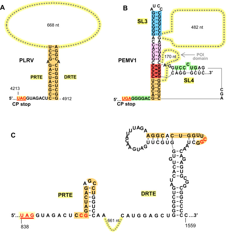
Fig 9. Comparison of PLRV and PEMV1 readthrough structures.
(A) Predicted readthrough structure of PLRV showing the key orange PRTE/DRTE interaction. (B) Proposed readthrough structure for PEMV1, including local and long-distance interactions. (C) Predicted local RNA secondary structures for PRTE and DRTE [13] regions in PLRV. Nucleotides shown in red were targeted for compensatory mutational analysis in Fig 8.

