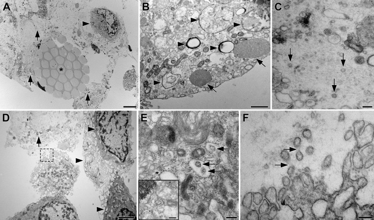
Fig. 1. TEM Images of clinical nasal swab specimens collected from a participant (subject 2), who tested positive for SARS-CoV-2 RNA by RT-qPCR.
A Cross-section through a swab fiber bundle (asterisk) with lysed (arrows) and intact cells (arrowheads); scale bar, 5 µm. B Cytoplasm of an intact epithelial cell with possible viral double membrane assembly structures (arrowheads) and cytoplasmic aggregates of unknown origin (arrows); scale bar, 1 µm. C Extracellular space with cytoplasmic material from lysed cells and three possible SARS-CoV-2 virions (arrows); scale bar, 200 nm. D Layer of intact epithelial cells with complex, interdigitating membrane protrusions (arrowheads) and lysed cell (arrow); scale bar, 2 µm. The cell in the center extends membrane protrusions into the lysed material outlined with a dashed box, shown at higher magnification in F. E Cytoplasm of an epithelial cell showing “outside-in” ribosomal structures (arrows)22 that are easily mistaken for virions, but are generated by budding of rER membranes into the lumen (insert); scale bar, 200 nm. F Plasma membrane of the boxed cell in D with convoluted membrane protrusions that are easily mistaken for virions, but can be identified as protrusions by faint connecting densities (arrows); scale bar, 200 nm.

