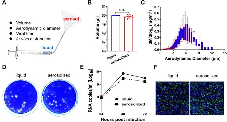
Figure 1.
Biological characteristics of Zika virus aerosolization. (A) Schematic diagram of the hand-held liquid aerosol pulmonary delivery device (HLAPDD). (B) Volume comparison between pre- and postaerosolization ZIKV. (C) Representative aerodynamic median mass diameter (MMAD) graph of Zika virus aerosolization by an aerodynamic particle size spectrometer. (D) The viral titres of pre- and postaerosolization ZIKV. Briefly, an HLAPDD was used to aerosolize ZIKV. The tip of the device was inserted into a 1.5-ml EP tube with a hole, and 200 μl of ZIKV aerosol was generated. The EP tube was left for 30 min and centrifuged for 30 s at 8000×g to collect the sample for the plaque assay. (E) Replication curve of pre- and postaerosolization ZIKV in BHK-21 cells. Briefly, BHK-21 cells were infected with liquid and aerosolized ZIKV, and cell supernatants were collected at 24, 48, and 72 hpi to detect the viral RNA load by the RT–qPCR assay. (F) Viral protein expression of pre- and postaerosolization ZIKV in BHK-21 cells. Briefly, BHK-21 cells were infected with liquid and aerosolized ZIKV, and then the cells at 48 hpi were fixed for immunofluorescence staining. The E protein was stained green, and DAPI (blue) staining indicated the nucleus. Scale bar: 100 μM.

