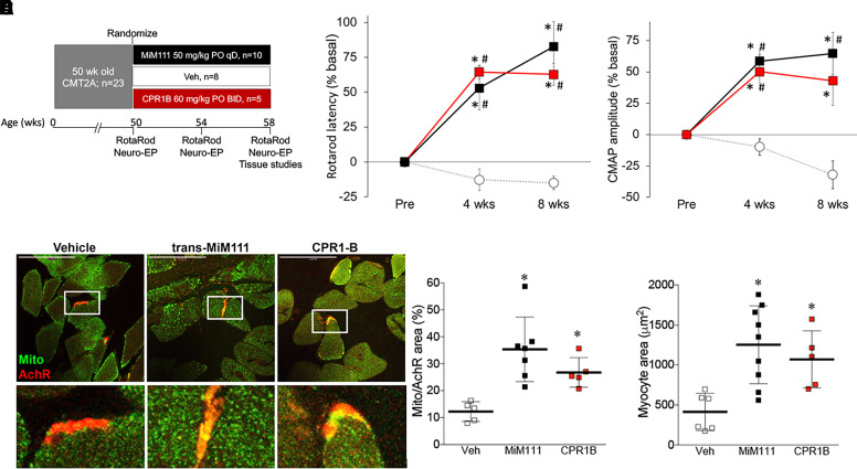
Fig. 4.
Sustained and burst mitofusin activation reverse neuromuscular degeneration in murine CMT2A. (A) Schematic depiction of experimental design. Once daily MiM111 confers transient mitofusin activation (Black); twice daily CPR1-B confers sustained mitofusin activation (red). (B, C) Improvement in neuromuscular function of 50-week-old MFN2 T105M CMT2A mice after mitofusin activation. (B) Rotarod latency; (C) hindlimb neuroelectrophysiological study CMAP amplitude. Results for each mouse are reported as % change in baseline. * = P < 0.05 versus baseline; # = P < 0.05 versus vehicle at same time. (D) Mitochondria (green, labeled with anti-COX IV) residency within acetylcholine receptors at neuromuscular synapses (AchR; red, labeled with Alexa Fluor conjugated α-bengarotoxin). Group quantitative data are to the right. (E). Tibialis muscle myocyte cross sectional area. (D, E) * = P < 0.05 versus vehicle (ANOVA); each marker represents a different mouse.

