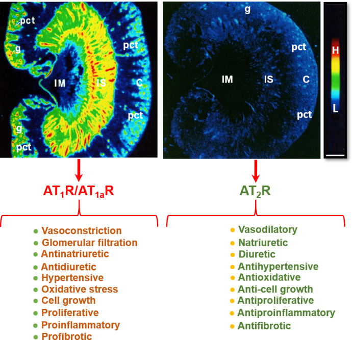
Fig. 5.
Localization of angiotensin II type 1 receptor (AT1R, largely representing its subtype a, AT1aR) and angiotensin II type 2 receptor (AT2R) in the rat kidney using quantitative in vitro autoradiography and opposing actions of AT1R/AT1aR and AT2R in the kidney. Panel A shows the anatomic localization of AT1R/AT1aR with high levels in the glomerulus (g) and the inner stripe of the outer medulla corresponding to vasa recta bundles, and moderate levels in the proximal convoluted tubules (pct) in the cortex (pct) and renomedullary interstitial cells (RMICs) in the inner stripe of the outer medulla between vasa recta bundles. The inner medulla (IM) expresses a very low level of AT1R/AT1aR. Panel B shows the anatomic localization of AT2R, with low levels in the outer cortex, corresponding to the glomeruli and the proximal tubules, and the inner stripe of the outer medulla, corresponding to vasa recta bundles and RMICs. Again, the IM expresses a very low level of AT2R. Red represents high level (H), whereas dark blue represents background levels (L). Modified from Zhuo et al. (1992, 1994).

