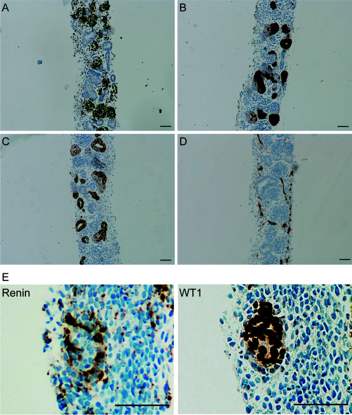
Fig. 8.
Immunohistochemical characterization of human induced pluripotent stem cell-derived kidney organoids. (A) Wilms’ tumor suppressor gene 1 (WT1) staining of glomerular structures; (B) Villin1 staining of proximal tubular structures; (C) E-cadherin staining of distal tubular structures; (D) CD31 staining of endothelial cells; (E) Renin staining, localized around WT1+ area. Organoids were generated as described by Shankar et al. (2021). Scale bar = 50 μm.

