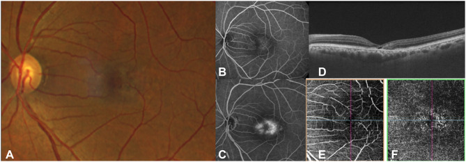
Figure 1.
Fundus photograph (A) and early (B) and late (C) fluorescein angiography, OCT (D) and OCTA (E and F) in a patient with MacTel. There is loss of retinal transparency temporal to fovea with angiographic leakage. (B and C) OCT (D) shows thinning of the parafoveal retina with hypo-reflective cavities in the outer retina. The OCTA (E and F) demonstrates dilation and telangiectasis of the deep capillary plexus temporal to fovea.

