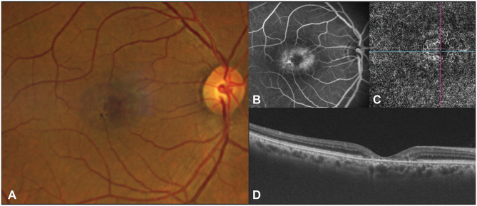
Figure 2.
Fundus photograph (A) showing blunted retinal venule and early pigmentation. The corresponding fluorescein angiogram (B) shows mild leakage in the late phase with hypofluorescence at areas of pigmentation. The OCTA (C) demonstrates dilation and telangiectasis of the deep capillary plexus temporal to fovea, and the OCT (D) shows foveal thinning with loss of the ellipsoid zone.

