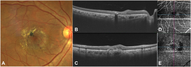
Figure 4.
Fundus photograph (A) depicts an active SNV with subretinal hemorrhage inferior to the fovea. Prominent crystalline deposits and pigment proliferation are seen temporal to fovea. The OCT images (horizontal and vertical cross-sections represented as (B and C) respectively) show sub-retinal hyper-reflective material with overlying retinal thickening and the OCTA (D and E) demonstrates a vascular network with a hypo-reflective flow void in the deep capillary plexus.

