FIG 3.
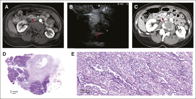
Example of a screen-detected stage IA pancreatic cancer (case 2). (A) Surveillance magnetic resonance imaging showing a new 1-cm hypoenhancing lesion in the head of the pancreas (arrow pointing to mass). (B) Confirmatory EUS showing a 1.5-cm hypoechoic lesion in the head of the pancreas without invasion of nearby vessels with cytology (not shown) diagnostic of a moderately differentiated adenocarcinoma. (C) Confirmatory computed tomography of the abdomen showing s 1.5-cm pancreatic head mass without upstream dilation or atrophy. (D) Whole-slide scanned image of a resected 1.4-cm lesion showing at 5× (E) a moderately differentiated invasive ductal adenocarcinoma confined to the pancreas. EUS, endoscopic ultrasound.

