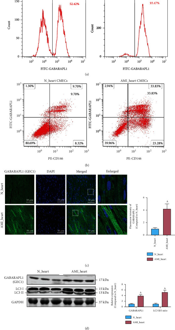
Figure 4.

The expression of GEC1 in the blood circulating endothelial cells (CECs) and heart cardiac microvascular endothelial cells (CMECs) after AMI. Flow cytometric analysis of the expression of GEC1 (a) in CECs and (b) in CMECs after AMI. (c) Immunofluorescence analysis of the expression of GEC1 in heart tissue after AMI (bar = 50 μm). (d) The LC3 II/I ratio and the protein expression of GEC1 in the heart of AMI rats. (a) p < 0.05 compared with the normal group.
