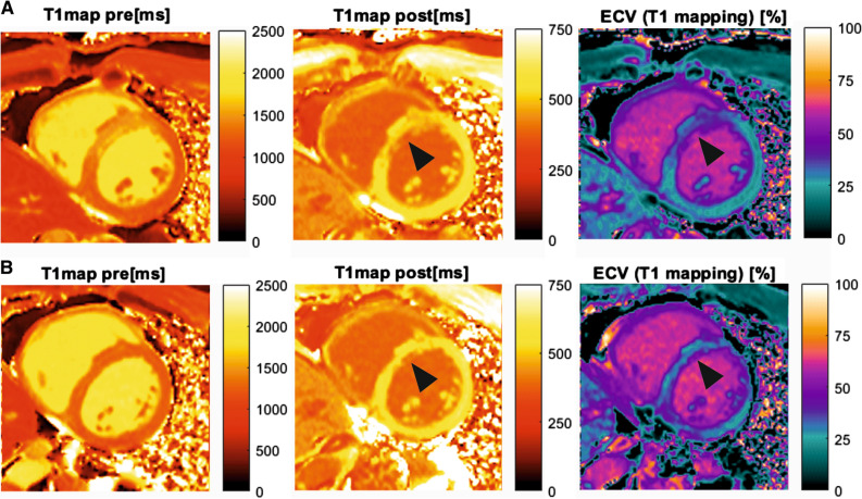
Figure 3.
CMR of patient after MI. CMR of the patient with acute MI. A T1- and ECV mapping performed during simultaneous PET/CMR examination following STEMI in LAD territory. The focal intense myocardial 68Ga-FAPI-04 uptake (Figure 2B) is concordant with the alteration in T1 and ECV mapping. B Control CMR of the same patient after six months. T1- and ECV mapping showed regressive edema of the myocardium in the previously infarcted area and improving T1 and ECV parameters. A small subendocardial scar remains in the (antero-)septal wall in a similar extent as the distinct areas of decreased post-contrast T1-times and increased ECV (arrowhead)

