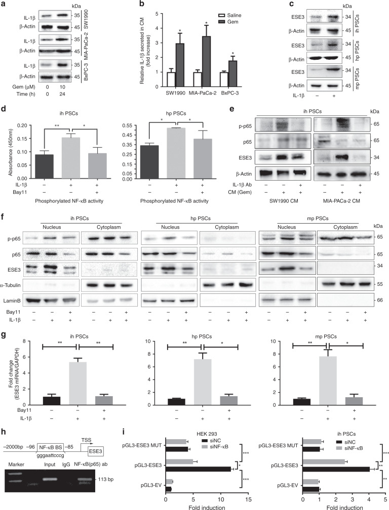
Fig. 5. Tumour-secreted IL-1β induced nuclear ESE3 (PSCs) expression by activating NF-κB.
a Western blot analysis of IL-1β expression in SW1990, MIA-PaCa-2 and BxPC-3 cell lines treated with Gem (10 μM) for 24 h. b ELISA of IL-1β secretion in the CMs of SW1990, MIA-PaCa-2 and BxPC-3 cell lines treated with Gem (10 μM) for 24 h. c Western blot analysis of ESE3 in ihPSCs, hpPSCs and mpPSCs treated with recombinant human IL-1β (100 ng/mL, 24 h). d Analysis of NF-κB (p65) transcriptional activities in ihPSCs and hpPSCs treated with recombinant human IL-1β (100 ng/mL, 24 h) and/or NF-κB (p65) inhibitor (Bay11, 8 μM; 12 h) by commercial kit. e Western blot analyses of p-p65 and ESE3 in ihPSCs cultured with CM (SW1990 and MIA-PaCa-2 cell lines treated with Gem) and/or IL-1β-neutralising antibody for 24 h. f Western blot analyses of p-p65 and ESE3 expression in nucleus and cytoplasm separation from the whole protein of PSCs treated with recombinant human IL-1β and/or Bay11. g RT-PCR analysis of ESE3 mRNA expression in ihPSCs, hpPSCs and mpPSCs treated with recombinant human IL-1β and/or Bay11. h Schematic of the structure of the ESE3 gene promoter. Shown is one κB-binding site and its location (upper). ChIP analysis of NF-κB binding to the ESE3 promoter in ihPSCs (lower). i Luciferase assay-based promoter activity analysis of HEK293 (left) and ihPSCs (right) knockdown NF-κB (sip65) and control cells (siNC) transfected with pGL3-ESE3, pGL3-Empty Vector (pGL3-EV) and pGL3-Mutation (pGL3-MUT). The cells were subjected to dual-luciferase analysis 48 h after transfection. The results are expressed as fold induction relative to that in the corresponding cells transfected with the control vector after the normalisation of firefly luciferase activity according to Renilla luciferase activity. The data are expressed as means ± SD from three independent experiments. *P < 0.05, **P < 0.01 and ***P < 0.001.

