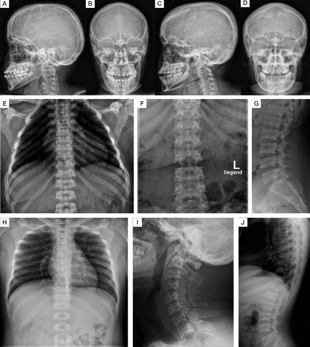
Figure 1.
Phenotype of skull, thorax and spine. Lateral skull radiographs show mildly thickened calvarias of S1 (A, B) and S2 (C–D). S1 and S2 show an open bite (A, C). Radiographs of chest and spine show broad (oar-shaped) ribs (S1 at 17 years (E, F) and S2 at 14 years (H)), broad vertebral bodies, platyspondyly and irregular endplates (S1 at 17 years (G), S3 at 18 years (J)). S1 shows a bell-shaped thorax (E). Radiographs of lateral spine additionally illustrate anterior beaking and irregular vertebral end plates (J). Lateral spine radiograph of the lumbar area shows platyspondyly with scalloping of the posterior end plates (S1 at 12 years (G)). Anterior inferior beaking of the cervical spine of S3 at 20 years (I). S1–S4, subjects 1–4.

