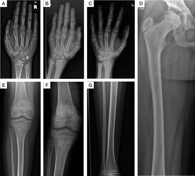
Figure 2.

Radiological abnormalities of the extremities. Hand radiographs demonstrate short third, fourth and fifth metacarpals, small carpal bones and small epiphyses of lower ends of radius and ulna (S2 at 14 years (A), S4 at 17 years (B), S3 at 18 years (C)). Posterior-Anterior (PA) projection hand radiograph of S2 at 14 years shows defective ossification of the carpal bones associated with retarded bone age. Apparent metaphysial irregularities and dysplasia along the metacarpophalangeal joints (A). PA right pelvis radiograph of S2 at 14 years (D) shows dysplasia and fragmentation of the capital femoral epiphysis associated with acetabular dysplasia. The shaft of the femur is overtubulated. Radiographs of the femora and tibiae demonstrate vertical striae in the metaphyses (S2 at 14 years (E), S4 at 17 years (F)). Tibia and fibula of S2 at 14 years show overtubulation of the shafts (G). Epi-metaphysial dysplasia of the inferior ends of the tibia and fibula. Metaphysial irregularities are associated with metaphysial striation (G). S1–S4, subjects 1–4.
