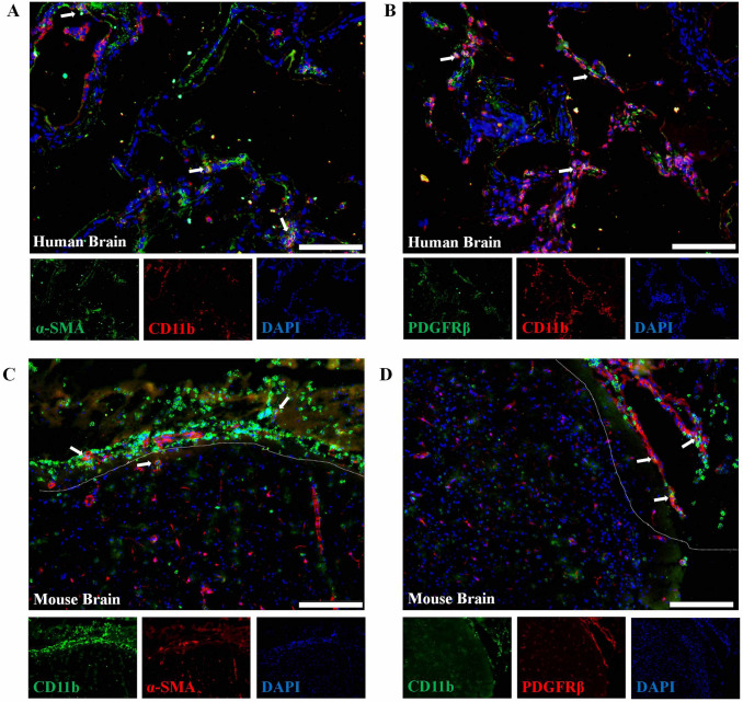
Fig. 1.
Immunofluorescence of PDGFRβ and CD11b in damaged brain tissue. A, B Immunostaining of the pericyte marker PDGFRβ/α-SMA (green) and CD11b (red) in brain tissue from a TBI patient. C, D Immunostaining of the pericyte marker PDGFRβ/α-SMA (red) and CD11b (green) in brain tissue from a TBI mouse. Damaged tissue is marked by dotted lines. Human and mouse brain tissues were collected within 24–48 h after TBI. Scale bars, 100 μm. Cell nucleus are stained with DAPI (blue).

