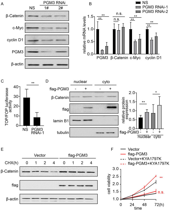
Figure 5.
Wnt/β-catenin signaling pathway is activated by PGM3. (A) Non-specific siRNA or PGM3 siRNA was transfected into HCT15 cells. 48 h after transfection, cells were collected, and western blotting was performed to detect β-catenin, c-Myc, and cyclin D1 protein levels. (B) Non-specific siRNA or PGM3 siRNA was transfected into HCT15 cells. 48 h after transfection, RNA was extracted and analyzed with real-time PCR. (C) The indicated siRNA and TOP/ FOP Flash reporter plasmid were co-transfected into HCT15 cells. 48 h later, luciferase activity was measured. (D) Empty vector or flag-PGM3 plasmid was transfected into SW480 cells. 24 h after transfection, β-catenin expression level in the nuclear and cytoplasm was assessed by western blotting. (E) PGM3 plasmid was transfected into SW480 cells, which were then treated with or without 50 μM CHX, western blotting was used to detect β-catenin level. (F) PGM3 plasmid was transfected into SW480 cells, which were then treated with or without 25μM KYA1797K, CCK-8 was used to examine cell proliferation. (A color version of this figure is available in the online journal.)

