
FIGURE 3.
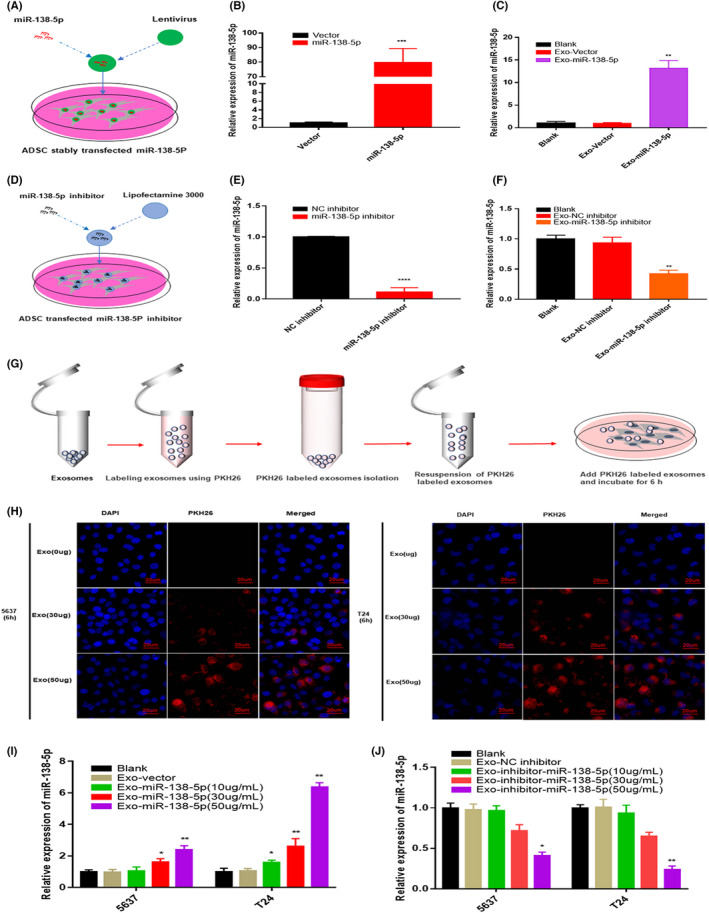
ADSCs could secrete exogenous miR‐138‐5p exosomes in a self‐assemble approach. (A) Construction of ADSCs stably expressing miR‐138‐5p with Lentivirus. (B) The relative expression level of miR‐138‐5p in ADSCs after stably transfected with miR‐138‐5p. U6 was used as internal reference. Data represent the mean ± SD. ***p < 0.001. (C) The relative expression level of miR‐138‐5p in exosomes after ADSCs stably overexpresses miR‐138‐5p. U6 was used as internal reference. Data represent the mean ± SD. **p < 0.01. (D) Transfection of miR‐138‐5p inhibitor into ADSCs by lipofectamine 3000. (E) The relative expression level of miR‐138‐5p in ADSCs after knockdown miR‐138‐5p. U6 was used as internal reference. Data represent the mean ± SD. ***p < 0.001. (F) The relative expression level of miR‐138‐5p in exosomes after ADSCs knockdown miR‐138‐5p. U6 was used as internal reference. Data represent the mean ± SD. **p < 0.01. (G) Schematic illustration shows that ADSC‐exo labeled with PKH26 and coculture with BC cells. (H) Exosomes internalization experiments showed that PKH26‐labeled exosomes (red) were taken up by BC cells. Nuclei were stained with DAPI. Scale bar, 20 μm. (I–J) The relative expression level of miR‐138‐5p in cocultured BC cells. U6 was used as internal reference. Data represent the mean ± SD. *p < 0.05, **p < 0.01