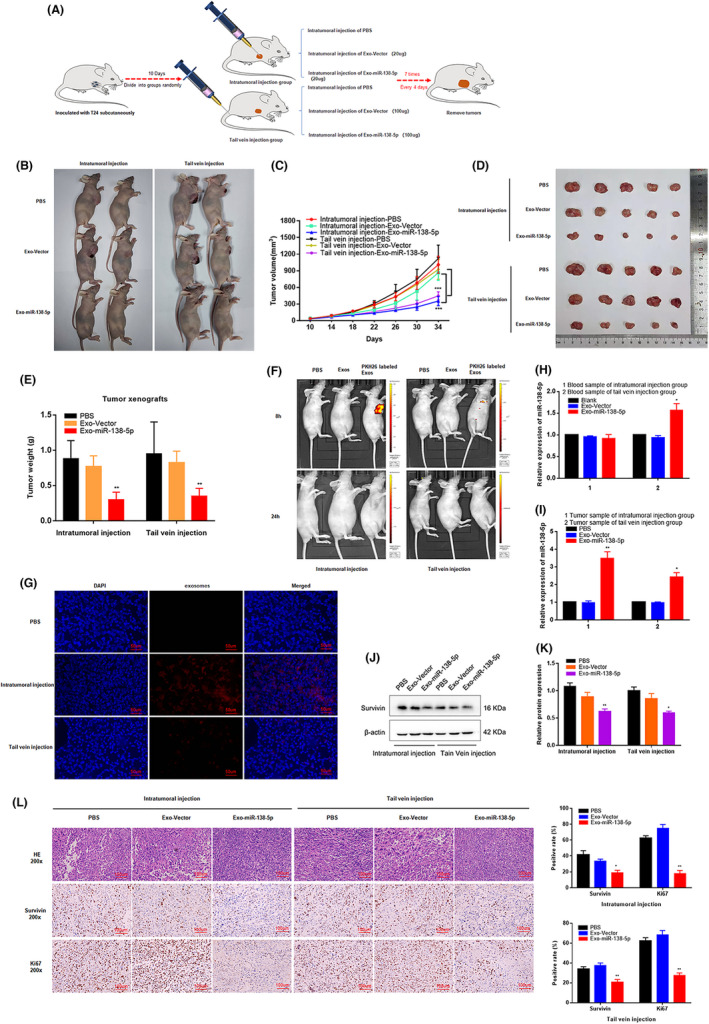
FIGURE 5.

ADSCs‐derived exosomes miR‐138‐5p inhibits BC growth in vivo. (A) Schematic representation of the establishment of the xenograft model (1 × 10⁷ cells per mice, n = 8 each group). (B–E) The growth rate and tumor weight significantly reduced in ADSCs‐exo miR‐138‐5p groups compared with the control groups. *p < 0.05, **p < 0.01, ***p < 0.001. (F) In vivo imaging system measured the delivery efficiency of exosomes in vivo. Fluorescence enrichment in tumor region after injection of PKH26‐labeled exosomes for 8 h. (G) Immunofluorescence was used to detect the distribution of exosomes in tumor tissues after injection of PKH26 labeled exosomes (red) 24 h. Scale bar, 50 μm. (H–I) The relative expression level of miR‐138‐5p in blood samples and tumor tissues of nude mice model. U6 was used as internal reference. Data represent the mean ± SD. *p < 0.05, **p < 0.01. (J, K) The expression level of survivin in tumor tissues was determined by western blot. β‐Actin used as internal reference. *p < 0.05. (L) Immunohistochemical representative images showed the survivin and ki67 positive cells in tumor tissue. Scale bar, 100 μm. *p < 0.05, **p < 0.01
