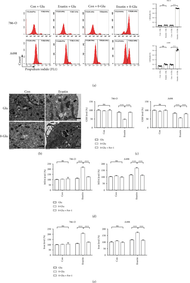
Figure 1.

Energy stress promotes ferroptotic cell death in renal cancer cells. (a) Cell death measurement shows 0-Glu promotes erastin-induced cell death. (b) TEM was used to observe morphological changes of cellular ultrastructure. Cells in 0-Glu and erastin treatment group present aberrant mitochondria. Black arrows indicate mitochondria. Scale bars: 1 μm. Independent experiments were repeated three times and representative data were shown. (c–e) 0-Glu and erastin treatment-induced GSH deletion, MDA generation, and cellular iron elevation are rescued by Fer-1. Glu: glucose concentration of RPMI 1640 medium. 0-Glu: 0 mM glucose. Data shown represent mean ± SD from at least three independent experiments. Comparisons were performed using Student's t-test. Fer-1: ferrostatin-1; ns: not significant. ∗∗∗p < 0.001.
