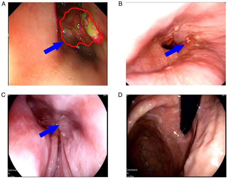
Figure 1.
Gastroscopic images of the patient in which the blue arrow indicates the tumor focus and the red outline represents the tumor focal boundary. (A) On January 11, 2019, a tumor lesion at the gastroesophageal junction blocked the esophageal lumen, preventing endoscopic exploration. (B) On May 22, 2019, the tumor was largely diminished and the endoscope was able to pass through the cardia. (C) Cardia and (D) fundus of the stomach were examined by gastroscopy on May 17, 2021, and no tumor tissue was visible with the naked eye.

