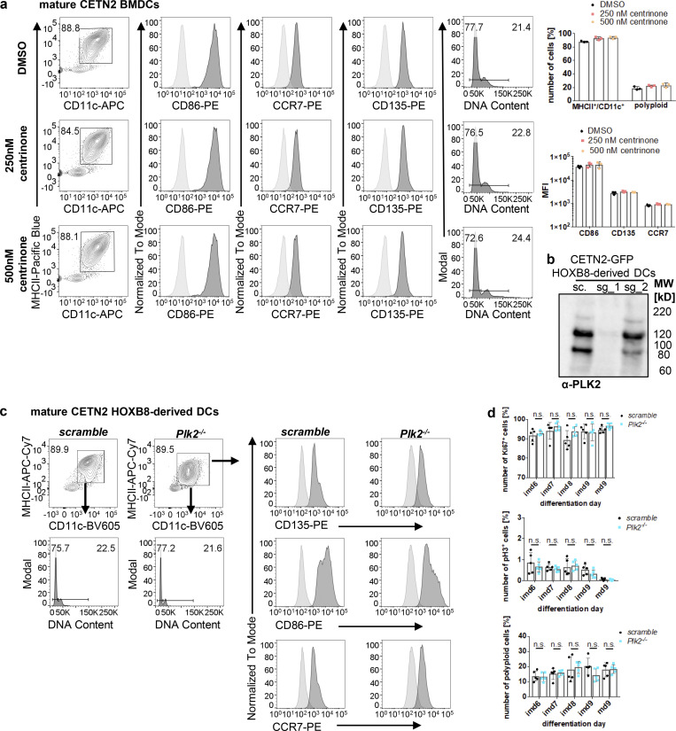
Figure S4.
PLK2 induction after LPS stimulation leads to untimely duplication of centrioles. (a) Differentiation and maturation of BMDCs in the presence of the PLK4 inhibitor Centrinone. Left: Mature DCs were identified as MHCII+/CD11c+ cells and further analyzed for DNA content and DC-specific cell surface marker. Black bars indicate gates for 2N and 4N cells. Unstained samples served as control and were included as light gray filled line. Staining for DC marker has been conducted in parallel with PE-conjugated antibodies. Right: Quantification of CD86, CCR7, CD135 in the presence or absence of Centrinone. Graphs show mean values ± SD of three independent experiments. N = 10,000 cells per experiment. MFI, mean fluorescence intensity. (b) PLK2 depletion in CETN2-GFP expressing HOXB8-derived DCs. Immunoblotting against PLK2 in control (sc., scrambled) and Plk2−/− (sg_1 and sg_2) DCs. Note that only single guide 1 (sg_1) and not sg_2 led to efficient Plk2 knockout. MW, mol wt. (c) Differentiation and maturation of HOXB8-derived Plk2−/− and control DCs. Mature DCs were identified as MHCII+/CD11c+ cells and further analyzed for DNA content (lower panels) and DC-specific cell surface marker (CD135, CD86, CCR7; right panels). Unstained samples served as control and were included as light gray filled line. Staining for DC marker has been conducted in parallel with PE-conjugated antibodies. Representative histograms of one out of three independent experiments are shown. N = 10,000 cells per experiment. (d) Quantification of proliferation markers (Ki67, pH3, and DNA content) in Plk2−/− (blue) and control (scramble; black) HOXB8-derived DCs. Graphs display mean values of ± SD of five independent experiments. N = 10,000 cells per experiment. n.s., non-significant (multiple, two tailed, unpaired t tests). Source data are available for this figure: SourceData FS4.

