FIG 1.
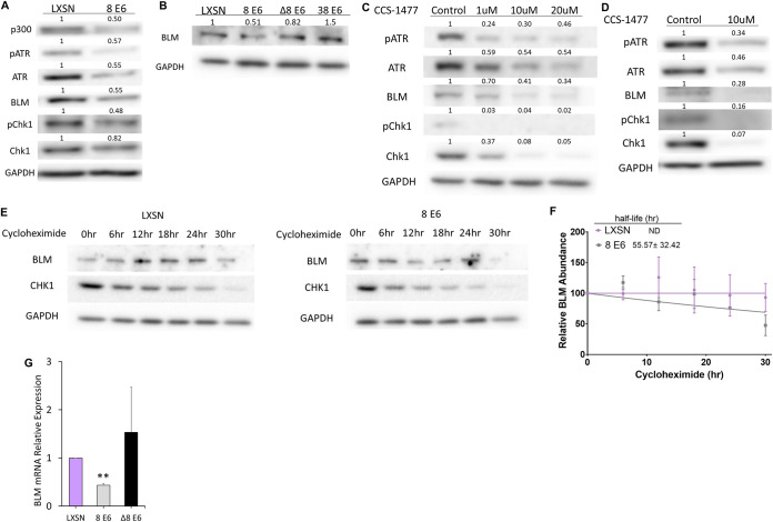
HPV8 E6 reduces BLM abundance. (A) Immunoblots of whole-cell lysates from iHFK cells expressing LXSN and 8 E6. (B) Immunoblots of whole-cell lysates HFK cells expressing LXSN and the indicated E6 genes. (C) Immunoblots of whole-cell lysates from iHFK LXSN cells treated with indicated amounts of CCS-1477 for 24 h. (D) Immunoblots of whole-cell lysates from HFK LXSN cells following 24 h of CCS-1477 treatment. (E) Representative immunoblots showing BLM abundance in iHFK cells at 0, 6, 12, 18, 24, and 30 h after 50-μg/mL cycloheximide treatment. (F) Average densitometry from three repeats of the experiment described in panel E. “ND” indicates that the half-life could not be determined by the indicated time points. BLM was quantified and normalized relative to GAPDH signal for each time point. (G) BLM mRNA expression in HFK cells measured by RT-qPCR and normalized to β-actin mRNA. Graphs depict means ± the standard errors of the mean from three independent experiments. Immunoblots are representative of at least three independent experiments. The numbers above the bands represent quantification by densitometry. Asterisks denote significant differences relative to LXSN (**, P ≤ 0.05 [Student t test]). In Δ8 E6, residues 132 to 136 were deleted from 8 E6.

