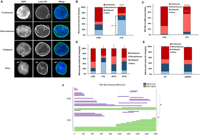
FIG 4.
HPV8 E6 reduces lamin B1 integrity of micronuclei. (A) Representative images of cells with intact (continuous and discontinuous) and disordered (collapsed and none) micronuclear envelopes. DNA is stained with DAPI (blue), and nuclear membranes were detected with antibodies against lamin B1 (green). A white arrow denotes the micronucleus in the “none” images. (B) Quantification of lamin B1 micronuclear morphology in asynchronous iHFK cells. n ≥ 234 micronuclei from three independent experiments. (C) Quantification of RFP-NLS-positive micronuclei segregated by micronuclear morphology (as determined by lamin B1 staining) in iHFK cells. (D) Quantification of lamin B1 micronuclear morphology in asynchronous HFK cells. n = at least 146 micronuclei from three experiments. (E) Depicts is the results from repeating the experiment as described in for panel D in HCT 116 cells. Quantification of lamin B1 micronuclear morphology in asynchronous iHFK cells. n ≥ 200 micronuclei from three independent experiments. (F) Length of time (in minutes) that RFP-NLS-positive micronuclei in iHFK RFP-NLS cells persisted across 30 h. Red, salmon, and blue asterisks denote a significant difference from LXSN corresponding with continuous, discontinuous, and no lamin B1 morphologies, respectively. Graphs depict means ± the standard errors of the mean from three independent experiments. Asterisks denote significant differences relative to LXSN (*, P ≤ 0.05; ***, P ≤ 0.001 [Student t test]). In Δ8 E6, residues 132 to 136 were deleted from 8 E6.

