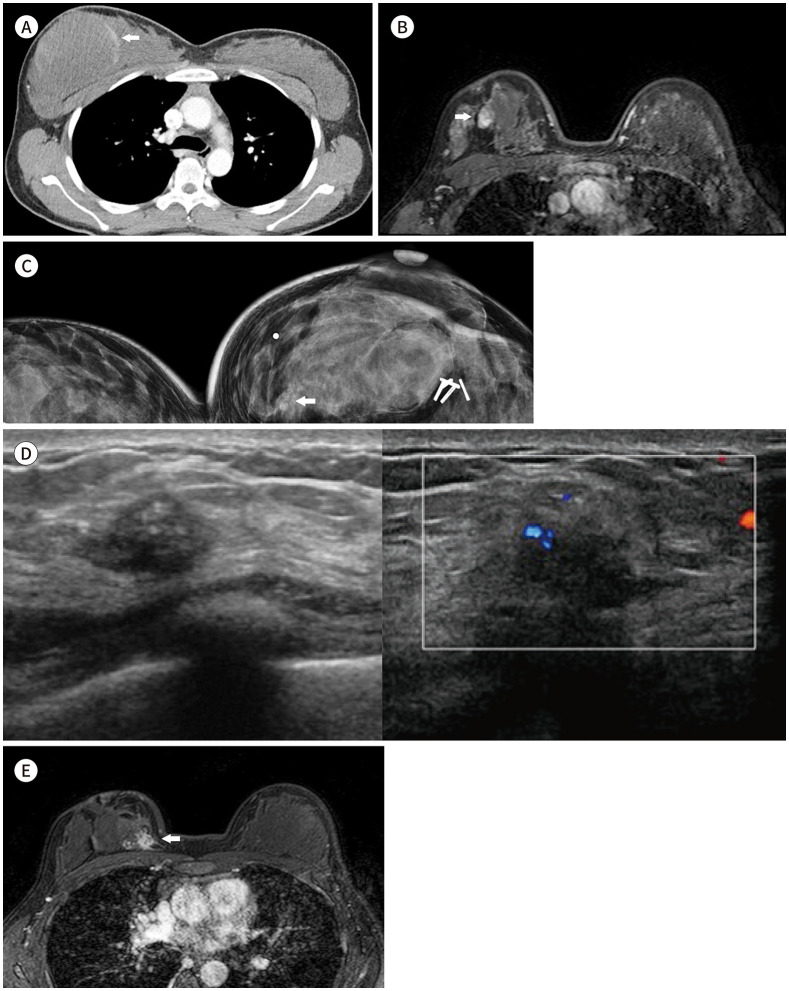
Fig. 1. A 29-year-old female with Li-Fraumeni syndrome and combined breast cancer.
A. Contrast-enhanced chest CT shows a 100 mm × 70 mm-sized heterogeneous low-density soft mass with a peripheral enhancing solid component (arrow) diagnosed as myxoid malignant fibrous histiocytoma (recently changed to pleomorphic undifferentiated sarcoma).
B. Dynamic contrast-enhanced MR with fat-suppressed T1-weighed gradient echo after wide excision of the mass shows a nearly 15-mm-sized irregularly shaped mass with heterogeneous enhancement in the right upper central breast (arrow), suggesting remnant mass.
C. 6 years after breast surgery shows focal asymmetry with grouped amorphous and pleomorphic calcifications (arrow) in the far inner portion of the right breast.
D. US image shows an irregular hypoechoic mass with internal echogenic dots corresponding to calcifications in the upper inner region of the right breast. Combined increased vascularity within the mass was demonstrated (transverse view and Doppler ultrasonography from the left side).
E. Dynamic contrast-enhanced T1-weighted fat-suppressed gradient-echo axial MR image shows a 26 mm × 18 mm-sized irregularly shaped mass with a spiculated margin and heterogeneous enhancement in the right upper inner breast (arrow).

