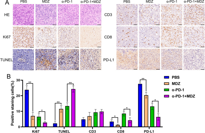
Fig. 5.
Immunohistochemistry showed that MDZ affected tumour growth and enhanced anti-PD-1 monoclonal antibody treatment efficacy in a xenograft mouse model. A The morphology of subcutaneous tumours in the four groups was confirmed by HE staining, as shown in the upper panel. Moreover, the upper panel indicates the results immunohistochemical staining for Ki67, TUNEL, PD-L1, CD3, and CD8 expression in the indicated groups. B The lower panel illustrates the statistical analysis of the levels of these indicators in the four groups. *P < 0.05, **P < 0.01

