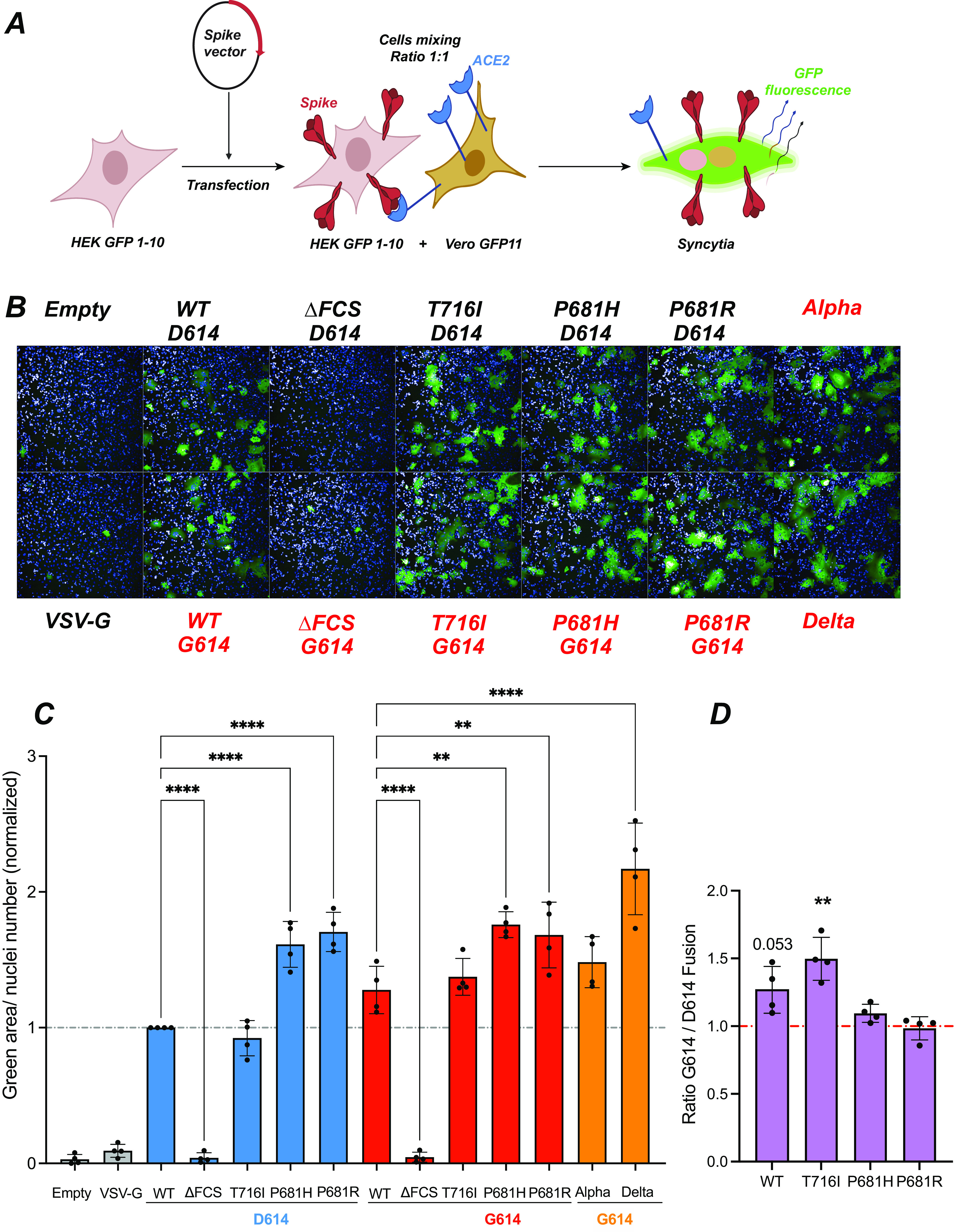
FIG 4.

The P681H/R mutations increase spike fusogenicity. (A) Principle of the cell-cell fusion assay. HEK GFP1-10 are transfected with spike vectors and then mixed with Vero GFP11 at a 1:1 ratio. Spike-ACE2 interactions lead to cell-cell fusion. Upon syncytium formation, complementation between the GFP1-10 and GFP11 fragments generates functional GFP, resulting in a fluorescent emission. (B) Representative images of a fusion assay monitored at 18 h after spike transfection. Cell nuclei were stained with DAPI (blue), while syncytia were detected by GFP fluorescent emission (green). The spike vectors used are shown, with those containing the D614G mutation in red. (C) Quantification of cell-cell fusion induced upon spike expression. The green fluorescent area divided by the number of nuclei is reported, with normalization to the WT spike condition. Statistics were measured by one-way ANOVA, with the Holm-Šidák correction for multiple comparisons. D614 backbone mutants were compared to the WT D614 spike, while G614 backbone mutants were compared to the WT G614 spike. (D) Ratios of cell-cell fusion measured for spikes in the G614 backbone to that in the D614 backbone. Statistics evaluating whether each ratio is different from 1 were measured by one-sample t tests. (C and D) Means and SD are shown for 4 independent experiments. Each dot represents the mean of technical triplicate measurements. **, P < 0.01; ****, P < 0.0001.
