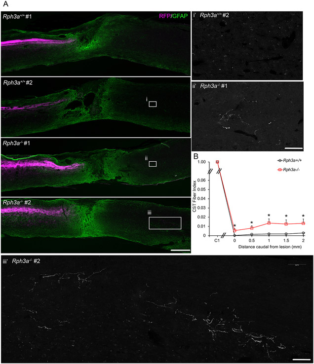
Fig. 10. Enhanced CST regeneration in Rph3a−/− mice after spinal contusion.
(A) Representative images of longitudinal section of lesion segment of spinal cord from the Rph3a−/− and WT mice with spinal contusion injury at T9 spinal level, stained with anti-RFP (magenta) and anti-GFAP (green) antibodies. Rostral is to the left and dorsal is up. Scale bar =500 μm. Boxed areas in i, ii and iii are magnified in i’, ii’ and iii’ for the RFP channel only. RFP-labeled CST fibers increased in the Rph3a−/− animals. Scale bar is 100 μm in i’, ii’ and iii’.
(B) Quantification of RFP-labeled CST axons in spinal cord caudal to lesion. RFP positive fibers are reported as a function of caudal distance relative to the SCI lesion site in the Rph3a−/− knockout group (n = 7) as compared to the WT group (n = 7). Data are mean ± SEM. *p < 0.05 (p = 0.023, F = 6.83, dF = 1), repeated-measure ANOVA for group effect across all sites, and *p < 0.05 (p = 0.028, F = 6.20, dF = 1; p = 0.032, F = 5.88, dF = 1; p = 0.027, F = 6.29, dF = 1; p = 0.044, F = 5.09, dF = 1; and p = 0.050, F = 4.74, dF = 1), significant difference between indicated pairs by one-way ANOVA.

