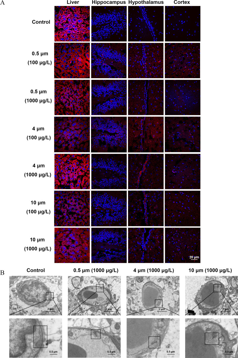
Figure 3.
The integrity of the blood–brain barrier (BBB) in mice treated with polystyrene microplastics (PS-MPs). Mice were exposed to three sizes of fluorescent PS-MPs dissolved in water for 180 days. (A) BBB integrity was detected by biotin tracer experiments. The liver and brain tissues were collected and immersed in paraformaldehyde, followed by a solution of 30% sucrose overnight. The tissues were embedded in OCT and cut into sections. The sections were stained with biotin (red) and DAPI (blue). The existence of biotin in brain tissues was examined by immunofluorescence microscopy ( mice/group, slides/mice). The biotin signal of the liver parenchyma was used as a positive control. Images of other mice are shown in Figure S2. (B) PS-MPs–induced ultrastructural changes in the BBB were detected by a transmission electron microscope. Upper images display the ultrastructure of BBB. Lower images refer to the magnified boxed areas. These are representative images ( mice/group). Images of other mice are shown in Figure S3. Note: DAPI, 4′,6-diamidino-2-phenylindole.

