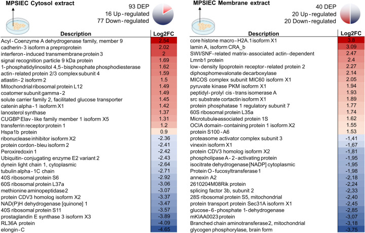
Fig 5. Differentially expressed proteins in MPSIEC after incubation with FhNEJ.
The top 15 proteins with highest (represented in red) or lowest (represented in blue) fold change within the MPSIEC membrane and cytosol extracts are shown. Icons created with BioRender.com.

