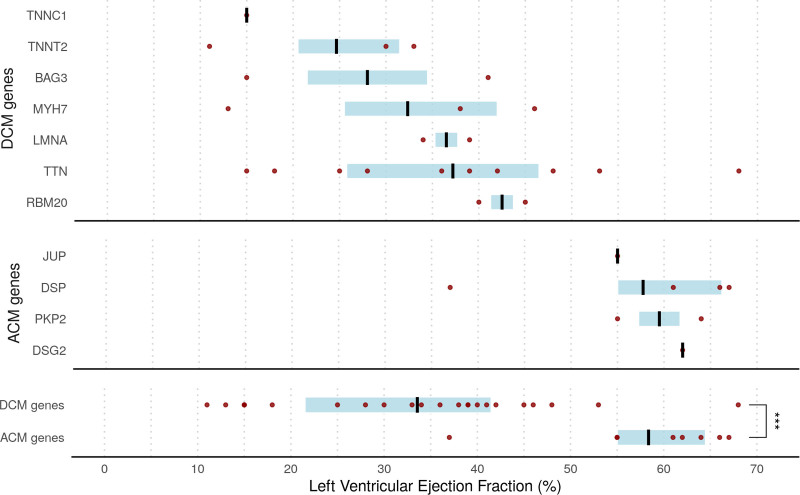
Figure 3.
Distribution of LVEF assessed by cardiac magnetic resonance in patients with acute myocarditis recruited in London (cohort 1; n=230) and Maastricht (cohort 4; n=106) stratified by presence of likely pathogenic variants in ACM- and DCM-associated genes. Black lines indicate median; blue shading shows interquartile range. Dots refer to individual patients. Note that genes with a single patient affected have left ventricular ejection fraction (LVEF) shown as an absolute value (applies to DSG2, JUP, and TNNC1). ACM indicates arrhythmogenic cardiomyopathy; and DCM, dilated cardiomyopathy.

