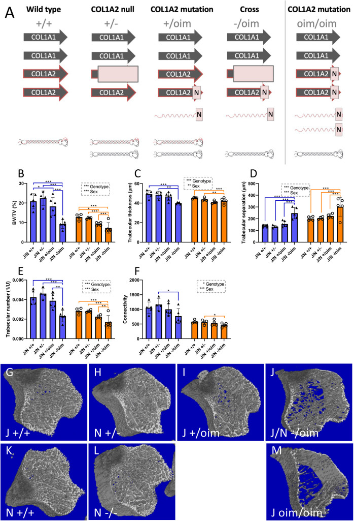
Fig. 8.
Bone structural properties are impaired in compound heterozygotes compared to heterozygotes of the oim or Col1a2 null lines. (A) Genetic differences between the heterozygous oim and Col1a2 null alleles and the compound heterozygous allele, with implications for collagen (I) protein synthesis. The homozygous oim allele is shown for comparison. Arrows indicate COL1 genes; N indicates mutation, light-red box indicates null allele. Folded heterotrimeric proteins are indicated in black and red; homotrimers are in black only. The presence of unincorporated mutant Col1a2 allele is indicated as a red waveform with a mutation (N). (B-F) μCT scans were performed on the knee joints of offspring from heterozygous crosses of each line. Reconstruction and analysis of scan files enabled determination of bone volume (B), trabecular thickness (C), trabecular separation (D), trabecular number (E) and connectivity (F). Blue bars/triangles, males; orange bars/circles, females. Blue (males) and orange (females) brackets show differences between genotypes. *P<0.05, **P<0.01 and ***P<0.001 (two-way ANOVA). n=4 for all groups, except male J/N +/+ and J/N -/oim of both sexes where n=5, and male J/N +/oim where n=6. Bars show mean±1 s.d. (G-M) Representative scan images from wild types from the oim line (G), Col1a2 null heterozygotes (H), oim heterozygotes (I), compound heterozygotes (J), wild types from the Col1a2 null line (K), Col1a2 null homozygotes (L) and oim homozygotes (M). Bone structural defects are more pronounced in compound heterozygotes than in oim heterozygotes.

