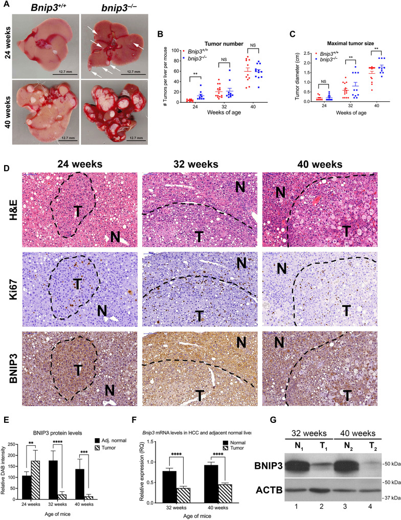
Fig. 1. Loss of BNIP3 promotes HCC initiation and tumor growth.
(A) Representative images of HCC tumors forming in the liver of Bnip3+/+ and bnip3−/− mice at 24 and 40 weeks of age following DEN injection. Scale bars, 12.7 mm. (B) Graph of tumor number forming in the liver of Bnip3+/+ (red) and bnip3−/− (blue) mice at 24 weeks (n = 9 per genotype), 32 weeks (n = 14 per genotype), and 40 weeks (n = 13 per genotype) of age following DEN injection. (NS, not significant; **P < 0.01). (C) Graph of tumor size in the liver of Bnip3+/+ (red) and bnip3−/− (blue) mice at 24 weeks (n = 9 per genotype), 32 weeks (n = 14 per genotype), and 40 weeks (n = 13 per genotype) of age following DEN injection (NS, not significant; ** P < 0.01). Scale bars, 100 μm (top left). (D) Liver sections from Bnip3+/+ mice at 24, 32, and 40 weeks of age following DEN injection, stained with H&E (top row), Ki67 (middle row), and BNIP3 (bottom row). (E) Quantification of immunohistochemical staining for BNIP3 on liver sections from Bnip3+/+ mice at 24 weeks (n = 5), 32 weeks (n = 7), and 40 weeks (n = 5) of age following DEN injection (**P < 0.05, ***P < 0.005, and ****P < 0.0005). (F) qPCR for Bnip3 mRNA isolated from HCC lesions and adjacent normal liver in Bnip3+/+ mice at 32 weeks (n = 12) and 40 weeks (n = 12) of age following DEN injection (****P < 0.0001). (G) Western blot for BNIP3 in protein lysates isolated from HCC tumor lesions (T) and adjacent normal (N) liver in Bnip3+/+ mice at 32 and 40 weeks of age following DEN injection.

