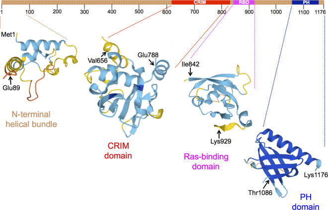
Figure 1. S. cerevisiae Avo1.
Top, schematized primary structure, highlighting locations of the conserved region in the middle (CRIM, red), Ras-binding domain (RBD, pink) and pleckstrin homology domain (PH, blue). Bottom, depiction of the three-dimensional structures of the indicated sequence segments, as predicted by AlphaFold2 [43]. Actual crystal structures for the C-terminal PH domains of Avo1 and mSIN1 have been determined [42], reflected in the dark blue coloring (the darker the shade of blue the higher the confidence level of the AlphaFold2 prediction).

