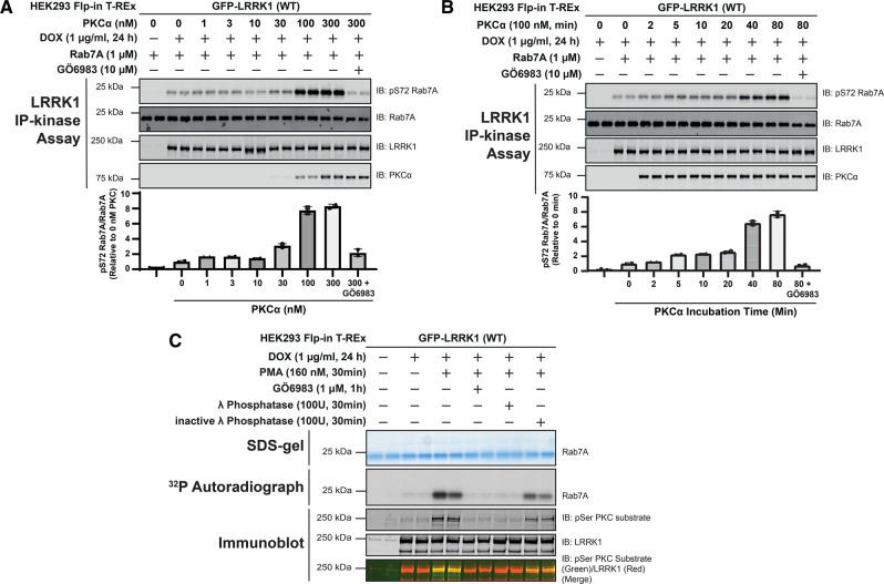
Figure 5. Time-course, dose-dependence, and reversibility of activation of LRRK1 by PKCα.
(A) HEK293 Flp-In T-REx cells stably expressing wild type (WT) GFP-LRRK1 were treated ± 1 µg/ml doxycycline for 24 h to induce GFP-LRRK1 expression. Cells were serum-starved for 16 h, lysed and GFP-LRRK1 immunoprecipitated and aliquoted as indicated. Each aliquot contains GFP-LRRK1 immunoprecipitated from 1 mg of HEK293 cell lysate. Step-1 of the LRRK1 kinase activation assay was setup by incubating GFP-LRRK1 aliquots with the indicated concentrations of PKCα and ±inhibitor GÖ6983 (10 µM) in the presence of non-radioactive MgATP for 30 min. PKC phosphorylated LRRK1 was then diluted 1.5-fold into a kinase assay containing recombinant Rab7A (1 µM) in the presence of non-radioactive MgATP (Step 2 of the kinase assay). Reactions were terminated after 30 min with SDS-sample buffer analysed by multiplexed immunoblot analysis using the LI-COR Odyssey CLx Western Blot imaging system with the indicated antibodies (upper panel). Combined immunoblotting data from two independent biological replicates (each performed in duplicate) are shown. Lower panel quantified immunoblotting data are presented as ratios of pRab7ASer72/total Rab7A (mean ± SEM) relative to levels observed with no PKCα added (given a value of 1.0). (B) As in (A) except in step 1 of the kinase activity assay 100 nM PKCα was incubated with immunoprecipitated GFP-LRRK1 for the times indicated. (C) As in (A) except that following serum starvation, cells were treated stimulated ± 160 nM phorbol 12-myristate 13-acetate (PMA) for 30 min and cells lysed. GFP-LRRK1 was immunoprecipitated from 1 mg of cell lysate incubated ± 100U of either active or 10 mM EDTA-inactivated λ phosphatase for 30 min. Following the phosphatase treatment, the beads were extensively washed and LRRK1 was subjected to kinase assay with recombinant Rab7A using Mg[γ-32P]ATP. Seventy-five percent of each reaction was subjected to SDS-polyacrylamide electrophoresis, stained by Coomassie blue (upper panel), and subjected to autoradiography (middle panel). The remaining reaction mixture was subjected to a multiplexed immunoblot analysis using the LI-COR Odyssey CLx Western Blot imaging system with the indicated antibodies (lower panels). Combined immunoblotting data from two independent biological replicates (each performed in duplicate) are shown.

