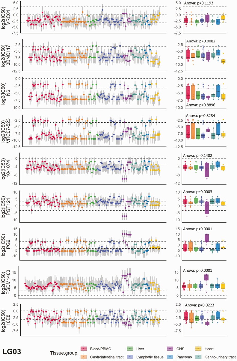
Figure 1.
The distribution of neutralization susceptibilities to the 9 selected broadly neutralizing antibodies (bNAbs) across all tissues; example is shown for participant LG03 in the Last Gift cohort. Each dot represents the predicted neutralization susceptibility with the respective prediction range (gray vertical lines) as calculated by our model. The dashed lines from bottom to top denote half-maximal inhibitory concentrations (IC50) = 0.1 µg/mL, IC50 = 1 µg/mL, and IC50 = 10 µg/mL. The box plots show the median values with the first and third quartiles across all tissue compartments for each bNAb. One-way ANOVA was used for across-group comparison. See also Supplementary Figure 3 for data on the remaining participants. Abbreviations: ANOVA, analysis of variance; CNS, central nervous system; PBMC, peripheral blood mononuclear cell.

