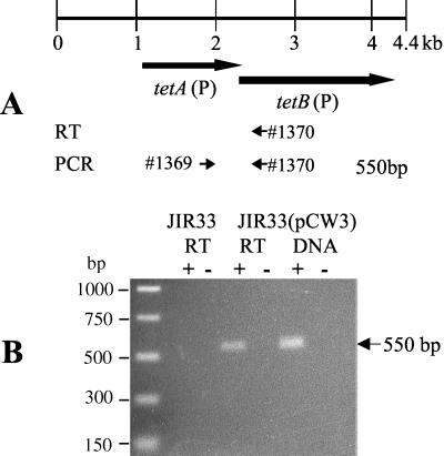
FIG. 1.
RT-PCR analysis of tet(P) RNA. (A) Schematic showing oligonucleotides utilized in RT-PCR experiments. The locations and extents of the tetA(P) and tetB(P) genes are shown. The locations of the oligonucleotide primers used in RT-PCR analysis are indicated by the numbered arrows. (B) Agarose gel electrophoresis of RT-PCR products. RT-PCR analysis was performed using total RNA that was isolated from JIR33(pCW3) cells grown in the presence of tetracycline (5 μg/ml) and from JIR33 grown without tetracycline. The positive controls, labeled DNA, used pCW3 templates extracted from JIR33(pCW3). The + and − labels refer to reactions performed in the presence and absence of RT, respectively. The + and − under the DNA label refer to PCRs performed in the presence and absence of DNA template, respectively.

