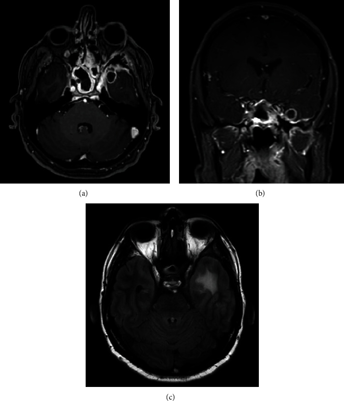
Figure 1.

(a) Axial T1-weighted brain MRI with contrast. (b) Coronal T1-weighted brain MRI with contrast. (c) Axial T2-weight fluid-attenuated inversion recovery brain MRI. (a, b) The images demonstrate a left medial temporal lobe well-defined, ring-enhancing lesion measuring 1.2 × 1.3 × 1.2 cm in anteroposterior, transverse, and craniocaudal dimensions. There is bilateral pachymeningeal enhancement, mostly involving the middle cranial fossa, worse on the left side. Asymmetric enhancement of the left cavernous sinus is noted as well as left eye proptosis. There is enhancement of the left lateral recuts muscle. There are significant mucosal thickening, enhancement, and air-fluid level in the sphenoid sinus and ethmoid sinus. (c) A left temporal vasogenic edema is noted. (a–c) These findings are suggestive of invasive sinusitis complicated by skull base osteomyelitis with intraorbital and intracranial extension.
