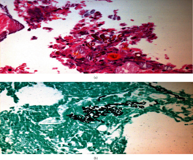
Figure 2.

(a) Hematoxylin-eosin (H&E) stain. (b) Grocott Methenamine Silver (GMS) stain. (a, b) Examination of the left temporal lesions revealed an abscess due to a fungal infection (branching nonseptate hyphae). The morphology of these hyphae is suggestive of Mucor species.
