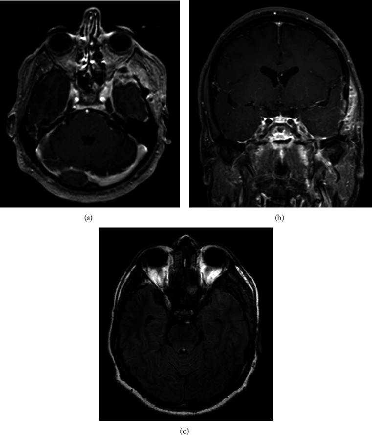
Figure 3.

(a) Axial T1-weighted brain MRI with contrast. (b) Coronal T1-weighted brain MRI with contrast. (c) Axial T2-weight fluid-attenuated inversion recovery brain MRI. (a, b) Follow-up images after 9 months of resection demonstrating persistent pachymeningeal enhancement in the left temporal lobe with no evidence abscess formation. (c) There is resolution of the vasogenic edema in the left temporal lobe.
