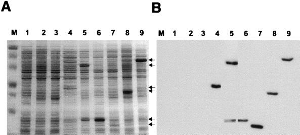
FIG. 4.
Detection of overexpressed His-tagged M. voltae flagellar accessory proteins FlaC, FlaD, FlaE, FlaF, FlaH, and FlaI derived from a T7 expression system. E. coli cultures harboring the respective flagellar accessory gene cloned into pET23a+ were grown to an optical density at 600 nm of 0.6 and then induced with 0.4 mM IPTG. Background vector controls were included in the analyses. Two hours postinduction, bacterial lysates (20 μg of total protein per lane) were analyzed by SDS-PAGE (12.5% gel) and the proteins were visualized by Coomassie blue staining. Arrows point to the gene-specific expressed proteins. (A) Lane M, molecular mass standards (in kilodaltons); lane 1, E. coli BL21(DE3)/pET23a+; lane 2, E. coli BL21(DE3)/pLysS/pET23a+; lane 3, E. coli BL21(DE3)/pKJ265/pET23a+; lane 4, E. coli BL21(DE3)/pKJ199 (FlaC); lane 5, E. coli BL21(DE3)/pLysS/pKJ198 (FlaD); lane 6, E. coli BL21(DE3)/pKJ194 (FlaE); lane 7, E. coli BL21(DE3)/pLysS/pKJ210 (FlaF); lane 8, E. coli BL21(DE3)/pLysS/pKJ200 (FlaH); lane 9, E. coli BL21(DE3)/pKJ265/pKJ266 (FlaI). (B) Immunoblot, developed with an anti-His monoclonal antibody, of a gel similar to that shown in panel A but containing 1/10 the amount of protein per lane.

