
Figure 3.
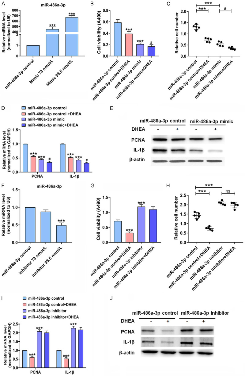
DHEA decreases VSMCs proliferation and inflammation by increasing miR-486a-3p expression. (A) VSMCs were transfected with miR-486a-3p control or mimic; qRT-PCR was used to detect miR-486a-3p. ***P < 0.001 vs. miR-486a-3p control group. (B-E) VSMCs were transfected with miR-486a-3p control or mimic for 24 h followed by stimulation with Ang II (10-7 mol/L) with or without DHEA (10-4 mol/L) for 24 h; MTS assay (B) was used to measure cell viability, and the Countess automated counter (C) was used for cell counting. The mRNA (D) and protein (E) expression of PCNA and IL-1β was measured. ***P < 0.001 vs. miR-486a-3p control group, #P < 0.05 vs. miR-486a-3p mimic group. (F) VSMCs were transfected with miR-486a-3p inhibitor or control for 24 h; qRT-PCR was performed to evaluate miR-486a-3p expression. ***P < 0.001 vs. miR-486a-3p control group. (G-J) VSMCs were transfected with miR-486a-3p inhibitor or control. After 24 h, VSMCs were incubated with Ang II (10-7 mol/L) with or without DHEA (10-4 mol/L). Cell viability assay (G), cell counting (H), qRT-PCR (I), and Western blotting (J) were performed. ***P < 0.001 vs. miR-486a-3p control group.