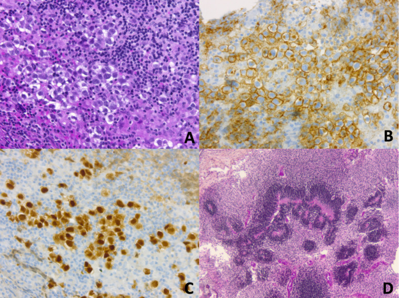
Figure 2.
(A–C) CNS Germinoma is composed of large round neoplastic cells, with clear cytoplasm, and high nuclear-to-cytoplasmic ratio. They are embedded in an inflammatory stroma of lymphocytes (mostly T cells) and histiocytes (A, Haematoxylin, Phloxine, Saffron (HPS) staining, original magnification (OM) x 200). Neoplastic cells demonstrate membranous and golgian expression of CD117/c-Kit (B, OM x 200). Nuclei are immunopositive for OCT4 (Octamer-binding transcription factor 4) (C, OM x 200). (D) CNS immature teratoma is characterized by a variable admixture of fetal and/or embryonic tissues derived from the three germ layers: ectoderm, mesoderm, and endoderm. Rosette-like structures resembling primitive neural tube are typical of immature neuroectodermal components (D, HPS staining, OM x 50).

