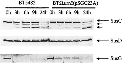
FIG. 4.
Immunoblots showing proteolytic sensitivities of SusC, SusD, and SusG in two strains, the wild type (BT5482) and the strain with the minimal starch utilization system [BTΩsusE(pSGC23A)]. Portions of a cell extract from each time point (100 μg) were loaded into each lane. The lanes are labeled according to the sampling time after addition of proteinase K. As expected, SusD was not degraded at all. This panel is provided to show that the outer membrane remained intact throughout the digestion period. Also, SusA, a periplasmic protein, was detected at the same concentration at all time points (data not shown). Electrophoresis conditions are described in Materials and Methods.

