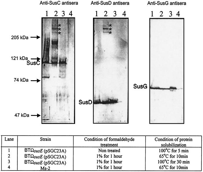
FIG. 5.
Immunoblots of the Sus outer membrane proteins of the minimal starch utilization system after formaldehyde cross-linking. Approximately 100 μg of protein from whole cells was loaded onto each lane. Sizes of molecular markers are given on the left. The Sus outer membrane proteins detected on the immunoblots are shown to the left of each immunoblot. Stars, cross-linked complexes. Electrophoresis conditions are described in Materials and Methods.

