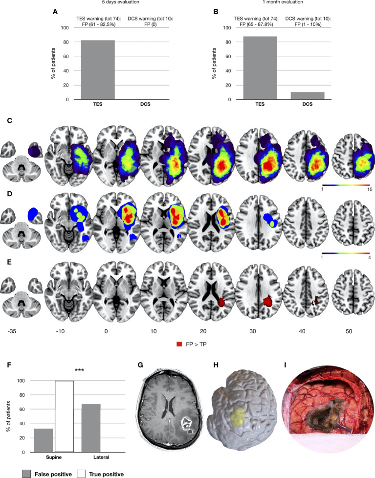
Figure 1.
Occurrence of false-positive results for TES and DCS considering the 5-day (A) and 1-month follow-up (B). Tumor volumes of patients with TES FP and TP results (1-month follow-up) are overlapped respectively in (C, D). The significant cluster predicting the occurrence of TES FP results is displayed in (E) (TFCE, p = 0.05). All tumor volumes, normalized to the MNI template, are visualized on left hemisphere axial slices. In (F) the distribution of supine and lateral craniotomies is reported between the FP and TP patients. Finally, in (G) (left panel, postcontrast T1-weighted images), a case of a high-grade glioma located in the left parietal lobe is presented in which a TES intraoperative MEP amplitude decreased but preserved postoperative motor status was recorded. In (H), a 3D render of the preoperative T1 is presented to show the lateral head positioning adopted for the resection. (I) A picture of the intraoperative field showing the brain shift and the presence of an air layer over the M1 convexity. ∗∗∗p < .001.

