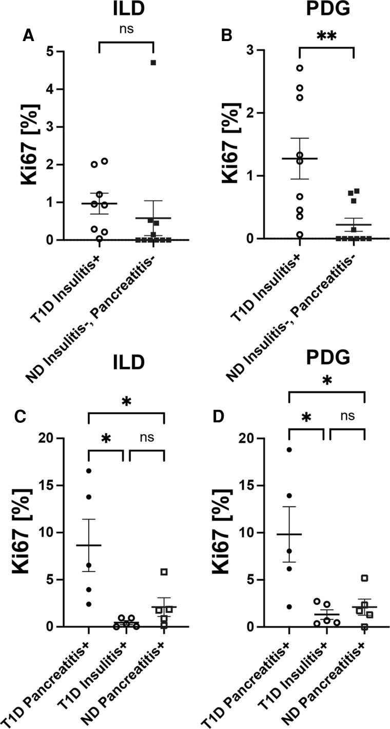
Figure 4.
Increased cellular proliferation in the ductal epithelium of type 1 diabetes (T1D) donors with insulitis or pancreatitis. Frequency of Ki67+ cells in (A) ILD and (B) PDG epithelium is higher in T1D insulitis+ subjects (black open circles, n = 10) compared with ND insulitis−, pancreatitis− subjects (blue filled squares, n = 10). Frequency of Ki67+ cells in the (C) ILD and (D) PDG epithelium of T1D pancreatitis+, insulitis− (black closed circles, n = 5) vs T1D, insulitis+, pancreatitis− subjects (black open circles, n = 5) vs ND pancreatitis+ subjects (blue open squares, n = 5). Each data point in (A) and (B) represents a specific insulitis+ section and matched control section evaluated per donor; see Table 1. Each data point in (C) and (D) represents an average of 1 to 3 sections evaluated per donor from the pancreatic head, body, and tail regions for T1D pancreatitis+ and ND pancreatitis+ donors and a specific insulitis+ section for T1D insulitis+ donors; see Table 1. (A-B) Data are presented as mean ± SEM with unpaired t test, P > 0.05. (C-D) Data are presented as mean ± SEM with one-way ANOVA with Tukey's post hoc test, * P < 0.05.

