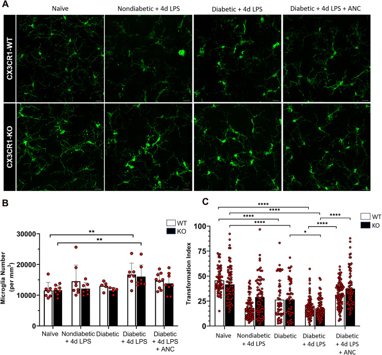
Figure 3.
Fibrinogen depletion reduces microglial activation in the diabetic murine retina. (A) Confocal images of IBA1+ microglia from 10-week nondiabetic and diabetic LPS-treated CX3CR1-WT and CX3CR1-KO mice show activated microglia with less ramification in comparison to highly ramified microglia with small soma in the naïve retina. Depletion of fibrinogen with ancrod shifts activated microglia towards increased ramification, similar to naïve microglia. (B) Quantification of microglial cell numbers indicates increased proliferation in the diabetic LPS-treated retina relative to the naïve CX3CR1-WT and CX3CR1-KO retina. Each point represents data from an individual mouse. (C) Morphometric analysis of IBA1+ microglia in the retinas of CX3CR1-WT and CX3CR1-KO mice by transformation index (TI) calculation reveals ramified microglia with higher TI values in the naïve and ancrod-treated diabetic retinas, and increasingly activated microglia with lower TI values in the nondiabetic and diabetic LPS-treated retinas, respectively. Each point represents the TI per individual cell. n = 5–9 mice per group. * P <0.05, ** P <0.01, *** P <0.001, **** P <0.0001. Scale bars: 25 µm.

