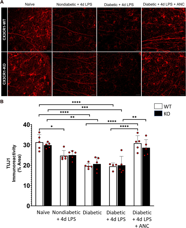
Figure 6.
Systemic fibrinogen depletion preserves axons in the diabetic retina. (A) Confocal images of retinas from 10-week nondiabetic and diabetic LPS-treated CX3CR1-WT and CX3CR1-KO mice show TUJ1+ axons (red) which are less dense in comparison to axons in the retinas of naïve and ancrod-treated diabetic mice. (B) Quantification of TUJ1 immunoreactivity in retinal tissues reveals decreased TUJ1+ area in the retinas of nondiabetic and diabetic LPS-treated CX3CR1-WT and CX3CR1-KO mice relative to naïve mice. Ancrod-treated diabetic CX3CR1-WT and CX3CR1-KO mice exhibit increased TUJ1+ axonal area compared to diabetic LPS-treated mice. n = 5–9 mice per group. Each point represents data from an individual mouse. * P <0.05, ** P <0.01, *** P <0.001, **** P <0.0001. Scale bars: 25 µm.

