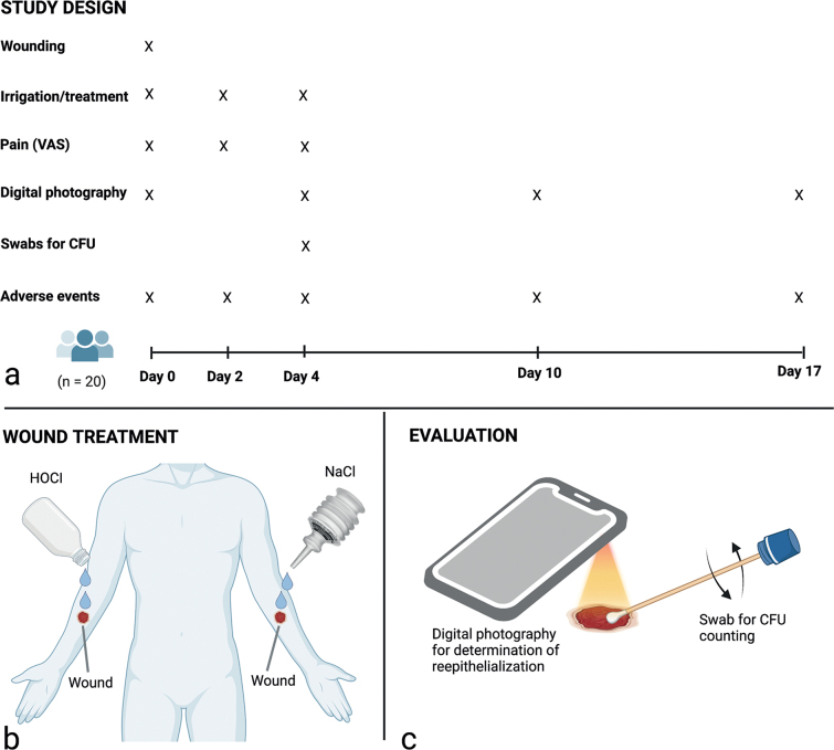
Fig. 1.
(a) Study design. (b) Treatments were randomized to the wound on the left or right forearm of the subjects, who served as their own controls. (c) Digital photographs were taken with an iPhone with an attached macro lens (days 0 and 4) or Handyscope (days 10 and 17) for blinded measurements of wound area and determination of re-epithelialization, and a sterile swab was used to quantitate colony-forming units (CFUs). Figure created with BioRender.com (Biorender, Toronto, ON, Canada).

